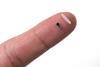
A self-powered, bioresorbable temporary pacemaker the size of a grain of rice has been developed by an international team of researchers. The device, which could potentially prevent lethal complications of heart surgery, could be especially valuable in young children.
After major cardiac surgery that requires stopping the heart, most patients receive a temporary pacemaker to ensure stable cardiac rhythm for several days or weeks. At present, this involves inserting thin leads from an external power source. These must then be extracted later. ‘The problem in many cases is that the pacing leads can be enveloped in scar tissue… The tearing of the scar tissue can sometimes lead to tearing of the normal, healthy tissue,’ explains biomedical engineer John Rogers of Northwestern University in Illinois, US. The resulting internal bleeding has led to many deaths, including the astronaut Neil Armstrong.
The new pacemaker, designed by Rogers and colleagues, requires no external leads to deliver the electrical pulse. Instead, 2mm-scale electrodes with different electronegativities implanted in the cardiac tissue form a battery, with the biofluids in the tissue as the electrolyte.
The team connected the electrode tips through a phototransistor. When this is not illuminated, the circuit remains open and the pacemaker is switched off. However, the researchers also mounted a rechargeable lithium-polymer battery onto the skin that continually measured electrocardiograms. If the heart monitor picked up an irregular heartbeat then the system could activate a pulsed LED emitting near-infrared wavelengths – to which tissue is relatively transparent. ‘That closes the circuit, leading to the delivery of electrical current to the surface of the heart that leads to the contraction of the cardiac muscle,’ says Rogers.
The entire pacemaker was non-toxic, small enough to be implanted through a 3mm needle and able to function for days or weeks – depending on which metals were used – before being almost totally broken down by the body in between one and three years, with the small remnant being biocompatible. Nothing in the device would interfere with an MRI either. Synchronised pacing of different regions of the heart – which is very difficult using conventional temporary pacemakers – could be achieved simply by using different wavelengths of light. Rogers believes that, thanks to its size, the device could be especially valuable in paediatric patients, who almost invariably require a temporary pacemaker after heart surgery.
The researchers are now focused on commercialisation. Rogers says five to seven years are normally required to achieve regulatory approval of an otherwise conventional new pacemaker, so this radical device will inevitably face strict scrutiny. ‘Having said that, we’ve done investigations on small and large animal models, and we’ve demonstrated the device on explanted human hearts from organ donors,’ he says. The team now intend to seek permission for trials in living humans.
‘This is an exciting paper,’ says Alex Abramson of Georgia Institute of Technology in the US. ‘There have been a lot of implants created that can be triggered via an external device – probably the most common one is an ultrasound-triggered device – but here they use light, which is a lot easier to generate than an ultrasound signal.’ He is especially impressed by the synchronised pacing of different locations. He has two caveats though: first, that the 40mm tissue-penetration depths achievable using light might be inadequate to reach the hearts of some patients, and secondly that the pacemakers’ lifetimes might be shortened considerably if frequent pacing were needed. Nevertheless, he says, ‘the stuff that they show is fantastic research’.
References
Y Zhang et al, Nature, 2025, DOI: 10.1038/s41586-025-08726-4
No comments yet