
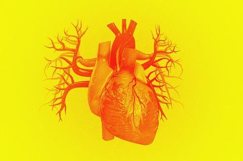
Nanotubes are being found in an increasing number of biological contexts, including the developing heart
Multicellular existence is collaborative. Our cells are in constant communication, checking out one another’s state and sending signals to modulate or moderate the behaviour of neighbours. The usual textbook story identifies three means of communication. Chemically specific signals emitted by one cell, such as hormones or neurotransmitters, might be registered by receptors on the surface of another; electrical signalling, like that among neurons, is common to most cells via channel proteins called gap junctions; and many cells will respond to mechanical signals using mechanosensitive ion channels. Cells can also send out packages called exosomes, lipid vesicles that bubble off from the cell membrane.
But for about 20 years we have known about another way for cells to get in touch: by forming nanoscale tunnels between the two. These so-called tunnelling nanotubes (TNTs) were first seen in rat cells in 2004.1 They are lipid tubules, typically around 20–500nm wide, that can bridge cells across distances of up to several cell diameters. Such nanotubes can wire groups of cells into complex networks linked by long, narrow corridors; occasionally the tubes may branch, each bridging more than two cells. All kinds of materials, including whole organelles such as mitochondria, can be exchanged between TNT-linked cells, undermining their identity as individual living systems. As one group of researchers has put it: ‘Ultimately, [TNTs] may challenge the cell theory formulated by Schleiden and Schwann … describing animal organisms as an assembly of individual, membrane-bounded entities.’2
TNTs typically emerge from a cell in an active process driven by the actin protein, which can polymerise into stiff rods that push on a membrane, rather as they do in the formation of the protrusions called filopodia that many cells use for migration, adhesion and tissue repair.3 The process seems to be guided by some sort of chemotaxis – by a chemical gradient between cells – since TNTs are generally linear protuberances that head right for their target cell. Once formed, a TNT can be stretched as cells move apart.
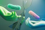
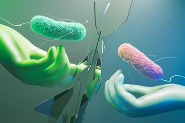
It has become ever more clear since TNTs were discovered that they have vital functions for cell signalling, for example in the immune system. There’s a dark side to them too. Viruses can pass from cell to cell this way, as can infectious prion proteins and other misfolded miscreants such as huntingtin (the protein responsible for Huntington’s disease).4 Even some bacteria can wriggle along larger TNTs, while bacteria themselves have their own versions of nanotubes. Cancer cells use them too: prostate cancer cells, for example, send vesicle traffic down TNT networks.5
Punching through
Two recent papers illustrate the diverse ways in which TNTs enable important biological processes. A team in the US has spotted TNT-like structures connecting mammalian heart muscle cells to endocardial cells (which form the heart lining) during the development of the heart.6 Powered by actin polymerisation, these tubes can punch their way from the heart muscle cells through the tangle of collagen and other fibrous proteins called cardiac jelly, so that the two cell types can exchange vesicles containing signalling molecules that guide the growth and patterning of heart tissue. Cardiac jelly is a kind of ‘soft skeleton’ that supports heart growth. Cells trying to communicate through this viscoelastic stuff could hardly rely on passive diffusion of vesicles or chemicals; creating tiny highways seems a much more reliable means of getting in touch.
Meanwhile, researchers at APstem Therapeutics and Stanford University in California, US, have found a remarkable way to exploit TNTs for engineering cell states.7 They wanted to solve a key problem for therapeutic uses of stem cells. The ability of such cells to form any tissue type (pluripotency) could be immensely valuable for regenerative medicine, but such versatility is all too apt to lead to tumour growth. So stem cells typically have to be made less ‘stemmy’ – partly or wholly differentiated – before being introduced to patients, which slightly defeats the object.
The researchers found that stem cells that retain developmental plasticity without being tumorigenic can be made by culturing them with ‘guide cells’ derived from human blood cells. It’s not yet fully clear why the guide cells have this influence – but they exert it by communicating with the stem cells (derived from human umbilical tissue) via TNTs, which seem to carry granules rich in RNA from the guide cells. The resulting ‘guide-integrated adult stem cells’ can regenerate skin and intestine in mice without causing tumours. Taking the tube is clearly sometimes the best way to travel.
References
1 A Rustom et al, Science, 2004, 303, 1007 (DOI: 10.1126/science.1093133)
2 H-H Gerdes, NV Bukoreshtliev and JFV Barroso, FEBS Lett., 2007, 581, 2194 (DOI: 10.1016/j.febslet.2007.03.071)
3 E Delage et al, Sci. Rep., 2013, 6, 39632 (DOI: 10.1038/srep39632)
4 M Costanzo et al, J. Cell Sci., 2013, 126, 3678 (DOI: 10.1242/jcs.126086)
5 C Vidulescu, S Clejan and KC O’Connor, J. Cell. Mol. Med., 2004, 8, 388 (DOI: 10.1111/j.1582-4934.2004.tb00328.x)
6 L Miao et al, Science, 2025, 387, eadd3417 (DOI: 10.1126/science.add3417)
7 S Li et al, Proc. Natl Acad. Sci. USA, 2025, 122, e2413043122 (DOI: 10.1073/pnas.2413043122)


No comments yet