A dye commonly found in food and cosmetics can be used to reversibly turn the surface tissues of a living mouse transparent. The novel technique, which the researchers call counterintuitive, requires no specialised equipment and allows the direct visualisation of anatomical features like muscle fibres, blood vessels and organs.
‘To make transparent mice in life is one of the dreams in our field,’ explains Hiroki Ueda, a synthetic biologist at Riken Centre for Biosystems Dynamics Research in Japan, who was not involved in the study.
Body tissue is opaque, in part, due to the varying refractive indexes of different components. Water-rich constituents, like cytosol, have low refractive indexes, while protein or lipid-based components have higher ones, with these mismatches causing light to scatter rather than penetrate tissue.
Existing methods for eliminating refractive index disparities can create transparent tissue. However, these kill living creatures as they use optical clearing agents (OCAs) that are toxic, such as tetrahydrofuran or acrylamide, and require the removal of molecules like lipids or water.
To investigate the possibility of achieving optical transparency in tissues using molecules that are strong absorbers of light, researchers from Stanford University, US modelled tartrazine and other similar molecules with promising optical properties. They found that water-soluble dyes could, theoretically, reduce the contrast in refractive indexes between water and lipids, reducing light scattering, turning tissues transparent.
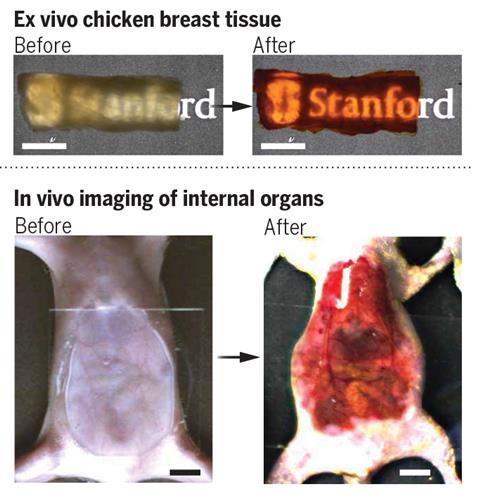
The team tested tartrazine, a water-soluble yellow azo dye already widely used in food, medicines and cosmetics, by dissolving it in an otherwise opaque suspension of colloidal silica. At 0.6M, the dye rendered the solution completely transparent in the red region of the visible spectrum, something that would take far higher concentrations of other OCAs like glycerol. The researchers further validated the technique by testing the dye on a muscle tissue model and thin slices of chicken breast.
Encouraged by the results, the researchers moved on to applying tartrazine topically to the scalp of a live, anaesthetised mouse. Using laser speckle contrast imaging, they were able to visualise the cerebral blood vessels in the mouse’s head, without removing the scalp as would normally be required.
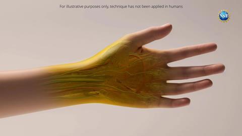
Rubbing tartrazine into the abdominal skin of a mouse similarly rendered it transparent in the red region of the visible spectrum, meaning its internal organs could be seen. Applying the dye topically to a mouse hindlimb let the researchers identify the periodic structures of sarcomeres, the contractile units of muscle fibres. By combining topical tartrazine with fluorescence microscopy imaging in mice engineered to express a bright red fluorescent protein in their cholinergic neurons, the researchers were able to visualise the movement of the middle part of the small intestine. In all cases, the transparency could be reversed by simply rinsing the skin with water.
Tests revealed that tartrazine diffuses through tissue at a much higher rate compared with other OCAs, creating transparency faster. As far lower concentrations of tartrazine are needed, there is also none of the shrinking and warping of tissues due to dehydration that occur with conventional OCAs.
Ueda says that the applications of this method for in vivo imaging of animals are numerous and immediate. However, he thinks that the application is, at least for now, likely ‘limited to probably animal research’.
‘Currently, this study has only been conducted on animals,’ explains Guosong Hong, a materials science and engineering researcher at Stanford, and one of the authors. ‘However, if the same technique could be applied to humans, it could offer a variety of benefits in biology, diagnostics and even cosmetics. Instead of invasive procedures, this technology could enable non-invasive methods for diagnosing conditions deep within the body.’ The team are now testing the safety and biocompatibility of their approach in human skin.
While the researchers’ models predict the possibility of identifying other molecules with long absorption wavelengths and sharp absorption peaks that could be even more efficient OCAs, Ueda feels that ‘to use [a] chemical or dye which is already approved to be a safe in human study for a long time and also [at a] higher dosage’ was ‘a great approach’ as it may help expedite use in humans.
References
Z Ou et al, Science, 2024, DOI: 10.1126/science.adm6869










No comments yet